|
|
|
| Ureteral calculi secondary to a gradually migrated acupuncture needle |
Masahiro Matsukia,*( ),Atsushi Wanifuchia,Ryuta Inouea,Fumiyasu Takeia,b,Yasuharu Kunishimaa ),Atsushi Wanifuchia,Ryuta Inouea,Fumiyasu Takeia,b,Yasuharu Kunishimaa
|
a Department of Urology, Hokkaido Social Work Association Obihiro Hospital, Obihiro, Japan
b Medical Incorporated Association Tenshunkai Tokachi Urological Clinic, Obihiro, Japan |
|
|
|
|
Abstract We herein presented a case of calculi secondary to a migrated acupuncture needle. A 74-year-old woman with a history of acupuncture therapy for lumbago was referred to our hospital for treatment of ureteral and renal pelvic calculi. Abdominal multi-detector computed tomography scans showed ipsilateral hydronephrosis and two calculi secondary to a migrated acupuncture needle. First, a percutaneous nephrolithotomy was performed to extract two calculi and fine needle fragments from the pelvis. Subsequently, residual needle fragments and calculi in the ureter were then removed by flexible transurethral lithotripsy using a holmium laser. In the present case, the formation of the calculi was caused by a migrated acupuncture needle. Calculi and needle fragments were removed safely endoscopically because the whole calculi and needle fragments were located in the ureteral lumen.
|
|
Received: 19 March 2018
Available online: 20 January 2021
|
|
Corresponding Authors:
Masahiro Matsuki
E-mail: mtkmatsuki@yahoo.co.jp
|
|
|
|
|
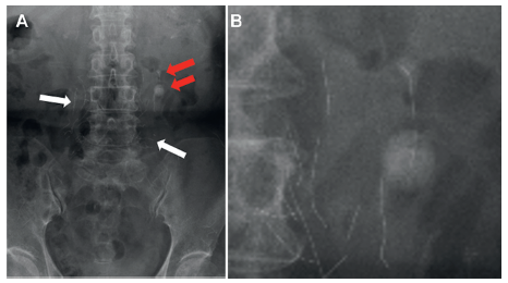
CT and X-ray results. (A) X-ray showing two calculi (red arrows) and many embedded needle fragments (white arrows) in the lumber region; (B) A larger image of the stones and needle fragments.
|

|
|
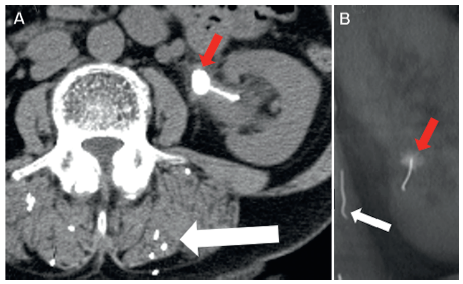
CT scan results. (A) Abdominal computed tomography (CT) scan taken 3 years prior showing calculus with the needle (red arrow) and many needle fragments (white arrow); (B) Coronal CT scan taken 5 years prior showing the faintly formed calculus with the needle (red arrow) and an embedded needle fragment (white arrow).
|

|
|
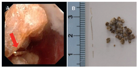
Flexible ureteroscope results. (A) Ureteroscopy showing the calculi fragments and a fine needle (red arrow); (B) Removed fine needle fragments and calculi.
|
| [1] |
Rempel S, Murti M, Buxton JA, Stephens W, Watterson M, Andonov A, et al. Outbreak of acute hepatitis B virus infection associated with exposure to acupuncture. Can Commun Dis Rep 2016; 42:169-72.
|
| [2] |
Domenicucci M, Marruzzo D, Pesce A, Raco A, Missori P. Acute spinal epidural hematoma after acupuncture: personal case and literature review. World Neurosurg 2017; 102:e11-4. https://doi.org/10.1016/j.wneu.2017.03.125.
|
| [3] |
Chun KJ, Lee SG, Son BS, Kim DH. Life-threatening cardiac tamponade: a rare complication of acupuncture. J Cardiothorac Surg 2014;31:61. https://doi.org/10.1186/1749-8090-9-61.
|
| [4] |
Lewek P, Lewek J, Kardas P. An acupuncture needle remaining in a lung for 17 years: case study and review. Acupunct Med 2012; 30:229-32.
|
| [5] |
Ernist E. Acupuncture: what does the most reliable evidence tell us? J pain Symptom Manage 2009; 37:709-14.
|
| [6] |
White A. [A cumulative review of the range and incidence of significant adverse events associated with acupuncture]. Acupunct Med 2004; 22:122-33.[Article in Japanese].
|
| [7] |
Yuzawa M, Hara Y, Kobayashi Y, Ishiyama S, Tozuka K, Nakamura S, et al. Foreign body stone of the ureter as a complication of acupuncture: report of a case. Hinyokika Kiyo 1991; 37:1323-7.
|
| [8] |
Minamida S, Iwamura M, Soh S, Sasamoto H, Ishikawa W, Kurosaka S, et al. [Spontaneous migration of a metal clip into renal pelvis after laparoscopic pyeloplasty: a case report]. Nihon Hinyokika Gakkai Zasshi 2007; 98:835-8.[Article in Japanese].
|
| [1] |
Guangju Ge,Zhenghui Wang,Mingchao Wang,Gonghui Li,Zuhao Xu,Yukun Wang,Shawpong Wan. Inadvertent insertion of nephrostomy tube into the renal vein following percutaneous nephrolithotomy: A case report and literature review[J]. Asian Journal of Urology, 2020, 7(1): 64-67. |
| [2] |
Rajeev Thekumpadam Puthenveetil, Debajit Baishya, Sasanka Barua, Debanga Sarma. Unusual case of nephrocutaneous fi stula -Our experience[J]. Asian Journal of Urology, 2016, 3(1): 56-58. |
|
|
|
|